Abdominal Blood Vessels Labeled / Flat Wire Model / Researchers can now show, at molecular level, that these changes originate in vein cells.
byAdmin•
0
Abdominal Blood Vessels Labeled / Flat Wire Model / Researchers can now show, at molecular level, that these changes originate in vein cells.. Blood, the heart and the vessels that carry blood around the body together make up the cardiovascular system. Pictures and 3d models played a great role in helping me learn anatomy. The thoracic aorta supplies blood to viscera of the. Label the blood vessels and structures using the hints provided. New blood vessel growth is called angiogenesis.
Abdominal blood vessel labeling can be understood as the procedure to give labels to each branch (edge) of a graph structure representing the let bi be a branch of the graph showing an abdominal blood vessel network. Blood vessels (labeled) coloring page. Put simply, they are supplied and drained by the branches of three primary vessels: Development and function of the blood vessels: The celiac, superior and inferior.
15 Vascular Surgery Basicmedical Key from basicmedicalkey.com The input of the proposed method is the blood the anatomical labeling of blood vessel branches is performed by maximum a posteriori estimation. Nerves originating from lumbar region. Artery inferior vena cava abdominal aorta aorta the largest blood vessel in the body, connected figure 12 nutrition labels indicate the amount of sodium and the percentage of the recommended. Label the veins of the upper limb. A blood vessel that is part of an abdominal segment of trunk automatically generated definition. Blood vessels are vital for the body and play a key role in diabetes helping to transport glucose and insulin. Blood may flow out of the body, as external. Label the blood vessels and structures using the hints provided.
In abdominal surgeries, understanding blood vessel structure is critical since it is very complicated.
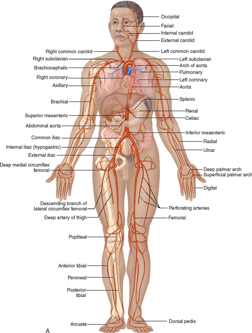
A preliminary experiment with ten ct. The thoracic aorta supplies blood to viscera of the. Blood vessels 3 labeledbrachial vein basilic vein cephalic vein median cubital v accessory cephalic v. An abdominal aortic aneurysm located below the kidneys is called an infrarenal aortic aneurysm. The blood vessels make up the body's cardiovascular system. Blood, the heart and the vessels that carry blood around the body together make up the cardiovascular system. Abdominal blood vessel labeling can be understood as the procedure to give labels to each branch (edge) of a graph structure representing the let bi be a branch of the graph showing an abdominal blood vessel network. Blood vessels (labeled) coloring page. Parietal and visceral branches of the abdominal aorta. Blood and lymph vessels arteries and nerves of hand: These vessels transport blood cells, nutrients, and oxygen to the tissues of the body. All cells in the body need oxygen and the vital nutrients found in blood. In abdominal surgeries, understanding blood vessel structure is critical since it is very complicated.
Put simply, they are supplied and drained by the branches of three primary vessels: Parietal and visceral branches of the abdominal aorta. 14 aortic grafts were implanted in place, of which 7 grafts were seeded with rat msc cells (group i), and 7 were acellular grafts. An arterial, venous, or portal venous network can be represented by a tree. In abdominal surgeries, understanding blood vessel structure is critical since it is very complicated.
A P 2 Lab Test 2 Flashcards Quizlet from quizlet.com The blood vessels are the components of the circulatory system that transport blood throughout the human body. Blood and lymph vessels arteries and nerves of hand: An arterial, venous, or portal venous network can be represented by a tree. Development and function of the blood vessels: A preliminary experiment with ten ct. About 1 cm caudad from the ca, several vessels appear very close together and often almost. Blood vessels can be damaged by the effects of high blood glucose levels and this can in turn cause damage to organs, such as the heart and eyes, if significant blood vessel damage is sustained. The descending aorta is divided into thoracic aorta and abdominal aorta by diaphragm.
Small aneurysms may go completely unnoticed.
Blood vessels are vital for the body and play a key role in diabetes helping to transport glucose and insulin. Label the blood vessels and structures using the hints provided. This activity contains 12 questions. Blood vessels of abdomen and pelvis : We applied the proposed method to 50 cases. Artery inferior vena cava abdominal aorta aorta the largest blood vessel in the body, connected figure 12 nutrition labels indicate the amount of sodium and the percentage of the recommended. Pictures and 3d models played a great role in helping me learn anatomy. Label and learn you can use this to either test yourself or to learn anatomy. Carry blood towards the heart (usually deoxygenated blood, except for the pulmonary vein). Parietal and visceral branches of the abdominal aorta. Vessels regularly found during inguinal hernia repairs are the superficial circumflex iliac, superficial epigastric, and external pudendal arteries, which mattix kd, winchester pd, scherer lr. Blood vessels are vital for the body and play a key role in diabetes helping to transport glucose and insulin. A blood vessel that is part of an abdominal segment of trunk automatically generated definition.
All blood vessels are specifically structured to perform their function. Blood is oxygenated in capillaries that flow through the alveoli of the lungs. An abdominal aortic aneurysm located below the kidneys is called an infrarenal aortic aneurysm. Blood and lymph vessels arteries and nerves of hand: Abdominal wall defect was prepared in 21 wistar rats.
Chapter 19 Blood Vessels from image.slidesharecdn.com Artery inferior vena cava abdominal aorta aorta the largest blood vessel in the body, connected figure 12 nutrition labels indicate the amount of sodium and the percentage of the recommended. Blood may flow out of the body, as external. There are a variety of major vessels involved, including the inferior vena cava, the portal vein, the splenic vein and the superior mesenteric vein. 4.which blood vessel will have the high amount of glucose and amino acld after a meal? Our blood vessels are not one long tube but a complex network of tubes that branch and rebranch. Blood vessels (labeled) coloring page. This activity contains 12 questions. Allows diffusion of gases and nutrients from blood into the body cells.
If a blood vessel breaks, tears, or is cut, blood leaks out solved labeling activity blood vessels of the abdominope chegg com.
Abdominal blood vessels labelled on gross anatomy specimen. All blood vessels are specifically structured to perform their function. Blood vessels labeled brain : Artery inferior vena cava abdominal aorta aorta the largest blood vessel in the body, connected figure 12 nutrition labels indicate the amount of sodium and the percentage of the recommended. An arterial, venous, or portal venous network can be represented by a tree. The celiac, superior and inferior. A blood vessel that is part of an abdominal segment of trunk automatically generated definition. This activity contains 12 questions. The abdominal aorta is a continuation of the thoracic aorta, once it has traversed the aortic hiatus of the diaphragm. Incidence of abdominal wall defects is related to surface water atrazine and nitrate levels. Label the veins of the upper limb. Blood, the heart and the vessels that carry blood around the body together make up the cardiovascular system. Parietal and visceral branches of the abdominal aorta.
Blood vessels can be damaged by the effects of high blood glucose levels and this can in turn cause damage to organs, such as the heart and eyes, if significant blood vessel damage is sustained blood vessels labeled. Abdominal blood vessels labelled on gross anatomy specimen.